


The lumbar osteocondrosis is a chronic degenerative-dystrophic disease of the lumbar spine that affects the structure of the intervertebral disks and a number of lumbar vertebrae.It affects people in predominantly working age.It manifests itself in various symptoms whose most important is pain in the crucifix and the legs, which limits movements in the crucifix.For diagnostics, research methods such as radiography, computed tomography or magnetic resonance are used tomography of the lumbar spine.In this article you can more detailed with the causes, symptoms and methods of diagnosing osteocondrosis of the lumbar spine.
Osteochondrosis is the result of aging of the body.These or other signs of this disease can be found in almost any person (!), From 25 years old.But here is the difficulty of these changes, the speed of their progression, the degree of clinical manifestations depends on many causes, primarily on how a healthy lifestyle leads a particular person.Moderate physical activity, mandatory morning gymnastics, the body's right position when performing a number of work (garden, construction, banal cleaning of the house and so on), the orthopedic mattress are the moments that prevent the development of osteochondrosis in the lumbar spine.
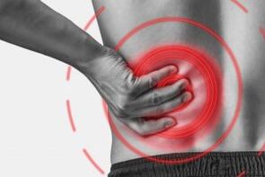
According to statistics, osteochondrosis of the spine in 80% of cases is the cause of back pain.
How does osteochondrosis develop?
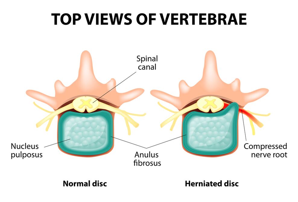
The entire spine consists of separate vertebrae, between the bodies of intervertebral disks.That is, between the two vertebrae is a disk.The disk consists of a gelatinous (pulpic) core and a fibrous ring.The nucleus contains a lot of water and gives depreciation and flexibility of the spine.The fibrous ring is located along the periphery of the jacket core, as if he is holding it inside himself.
With a prolonged increased load on the pier, it changes its physiological properties, loses water and dries and eventually sequences: The disk is flattened and the spines come close to each other.Along with such processes, in the core's jacket, the fibrous ring loses its elasticity and begins under the influence of mechanical loads to protrude.This is called projections.Then the fibrous ring falls and a gelatinous core falls through the resulting holes: a hernia of the disk occurs.A plot with two adjacent vertebrae and a disk located between them, called the back segment, acquires excess mobility, thereby increasing the load on the nearby segments.The overload of the nearby segments triggers a similar pathological process in them.These changes are called osteochondrosis.
In order to somehow secure the spine somehow, bone growth is formed along the edges of the spines, which increases the area of support.This phenomenon is called spondylosis.Changes in the joints between vertebrae are called spondylo -arthrosis.Usually all three pathologies - osteocondrosis, spondylosis, spondy arthritis - go nearby.
Causes
Why does osteochondrosis occur?To date, there are several theories about the occurrence:
- Mechanical theory: Perhaps the main cause should be considered as a regular increased load on the spine.Therefore, osteocondrosis is an almost mandatory fate for movements, miners, builders and people in such professions.The occurrence of osteocondrosis in the lumbar area is mainly associated with the slopes and lifting of severity, forcing an unpleasant working position;
- Another factor in development is the wrong attitude that is in the wrong position, which is especially relevant to mental workers;
- Sometimes the role of hereditary features is played by the spine and nutrition of its individual structures;
- Traumatic theory: Any trauma to the spine (even the most insignificant) is capable of starting a degenerative process;
- Hormonal metabolic disorders and endocrine diseases can adversely affect metabolism in the tissue of the spine and contribute to the development of osteocondrosis;
- Age theory involves the natural wear of disks in the process of life.
Rarely can only one of these theories explain the occurrence of osteochondrosis in each case.More often at the same time, several factors are "to blame".
In the occurrence of osteocondrosis of the lumbar spine, overweight plays an important role, as it is in itself an overload for the spine.The higher the body mass index (degree of obesity), the more significant changes in the spine is normal.Among other reasons provosing the appearance of osteocondrosis, one may notice:
- sedentary lifestyle;
- IMorly nutrition (fast food, excess sweet, semi -finished products: all this leads to an imbalance of trace elements) and lack of fluid;
- Anomalies of the spine (for example, the presence of a further lumbar vertebrae);
- constantly wearing tall -te -heeled shoes;
- pregnancy (due to excess load on the lumbar spine);
- Sudden ending of training in people who are professionally involved in sports;
- Smoking and abuse of alcohol: as factors that accelerate the aging process in the body.
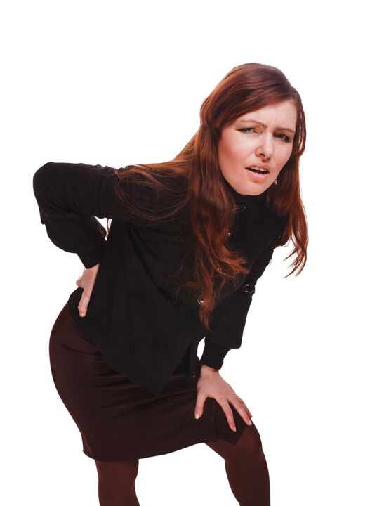
Symptoms
The most important manifestation of osteocondrosis in the lumbar spine is pain.The nature of the pain, the place of the occurrence and the direction of distribution depends on which receptors are irritated, that is, how gross changes in the disk and the surrounding tissue that are projections or hernia, in which direction the projection was formed and so on.
Reflex and compression syndromes are separated with osteochondrosis of the lumbar spine.
Reflex syndromes develop in cases where the receptors of the fibrous ring of the affected disk, ligaments and joint capsules located nearby are irritated.They are reflexive because in addition to pain is accompanied by muscle-tonic, vegetative-vascular or neurodistrophic reflex changes, that is, irritation with reflexes is transmitted to other structures, causing symptoms mainly from the side of the soft tissue.
Compression syndromes occur as a result of compression (compression) of nerve roots, blood vessels or spinal cord formed by osteocondrosis in changes.

Reflex -Syndromes in the lumbar spine
Lumbago(Feeling): Acute sudden pain in the crucifix that occurs with an awkward movement or at the time of physical excitement (much less often - without any obvious reason).It is believed that the occurrence of lumbago is associated with the movement of a jacket core inside the fibrous ring, that is, it develops in the initial stages of osteocondrosis.Often the pain is described as "feeling", "the rod was stuck in the crucifix."Patients freeze in the position where pain caught them.The slightest feature is caused by an increase in pain (sneezing, coughing, an attempt to turn in bed, move your foot).If a person was in a prone position at the time of the development of lumbago (most often happening), he cannot fix.A pronounced muscle tension in the lumbar spine seems reflexive.Along the vertebrae in this area is felt a muscle roll, which is sometimes visible to the naked eye without touch, and muscle tension is so pronounced.Feeling painful to the patient.Such an increased muscle tone performs an immobilizing role and protects the affected lumbar spine against pathological mobility, which can provoke a deterioration in the state.The natural bending of the spine in the crucifix (lordosis) is flattened, perhaps curvature (scoliosis) is possible due to muscle tension.
Lumbalgia- Another reflective syndrome at the lumbar level.This expression also means the presence of pain in the lumbar area.But unlike lumbago, the pain does not occur acute, but gradually within hours or even days.The pain is stupid, moderate intensity, intensifies during movements, in a sitting or standing position when moving from one position to another.A slight relief brings the position like lying or back with a roll under the crucifix, but the passive rise of the directed leg in this position causes increased pain in the crucifix (Lassa symptom).Palpation of the lumbar spine is painful, but the reflex voltage of the muscle is less pronounced than with lumbago and sometimes absent.Movements in the lumbar spine are limited but possible.This means that the patient can bend down and to the sides to a particular level (and then intensify the pain).
Sciatica- Another selection of reflective syndrome at the lumbar level.By this expression is meant pain in the crucifixion that gives the back and leg (on the back).The pain is different, mostly sore, but can be regularly intensified by the type of "fireplace" in the leg.As with lumbalgia, it intensifies with any movements, walking, strenuous, falling in the back.The symptom of Lassa is usually positive.Palpation of the lumbar spine is painful as well as pressing some points (for example, in the middle of the line separating the buttocks from the thigh, in the middle of the back of the thigh, in the middle of the popliteal fossa).There are tension in the crucifix.The slopes forward and until the sides are limited.
Compression syndromes in the lumbar spine
The clinical feature depends on the structure of compression.
Between the vertebrae of each intervertebral hole are nerve roots (spinal cord nerves): left and right.If pathological formations to osteocondrosis of the lumbar spine (mainly slices of disks) squeeze the roots, radiculopathy is developed whose symptoms are different for each root.Common to all radiculopathies in the lumbar area is the increase in pain during sneezing, coughing, movement in the crucifix (especially lashes forward), the presence of muscle tension in the crucifix, restriction of movements in the lumbar spine.The following types of radiculopathies of the lumbar spine are most common:
- Radiculopathy L1, L2, L3: Pain occurs in the crucifix, gives to the expected thigh.In the same area, the occurrence of paresthesia (a feeling of searching goosebumps, numbness) is possible, superficial sensitivity is disturbed (an acute touch from the usual is not separated, the feeling of cold and hot) is lost.The knee reflex falls, the weakness of quadriceps in the thigh is revealed;
- Radiculopathy L4: The pain from the crucifix gives the anterior part of the thigh, the inner surface of the knee joint and slightly lower along the inner surface of the lower leg.In the same areas, paresthesia is felt and the surface sensitivity is lost (reduced).Weakness of the quadriceps muscles in the thigh also develops, the knee reflex falls;
- Radiculopathy L5: One of the frequent locations.The pain gives the back along the outer edge of the thigh along the front surface of the lower leg to the inner edge of the foot and thumb.Paresthesia is felt here, superficial sensitivity is disturbed, and a pain is given here when sneezing and coughing.In addition, there are difficulties in extension of the thumb of the foot, as the muscle performing this action is inner wing by the kin L5.It is sometimes difficult to stand on a heel with an exposed foot;
- S1 radiculopathy is also often found with osteocondrosis of the lumbar spine.The pain gives the back along the outer edge of the thigh along the outer edge of the lower leg to the outer edge of the foot and the 5th finger, heels.These zones are characterized by a sense of paresthesia, a decrease in surface sensitivity.Achilles reflex is reduced.With the damage to this spine, the weakness of the muscles of the lower leg and flexors is developed in the foot, making it difficult to stand and walk on the socks.
The simultaneous development of radiculopathies of several roots is possible, this is especially characteristic of L5, S1.It happens that a hernia is pushing more roots.
If the slice's flock clings back, it can squeeze the spinal cord.This is only possible when the hernia is located in the upper reference point, as there are no spinal cord swirls under II -river virtues (the spinal cord roots are exposed to compression and the ponytail -syndrome develops).
If the vessels in the lumbar area are exposed to having the pressure that performs blood flow to the spinal cord develops in the event of an acute circulation disorder, a spinal cord and with prolonged compression - myelopathy.Myellopathy is manifested by bilateral weakness in the muscles of the legs, starting from the foot and gradually going up.The sensitivity of the legs is disturbed, Achilles reflex is lost and later knees.It is possible to appear urination disorders (frequent, "imperative" craving that requires immediate satisfaction, urinary incontinence).
Diagnostic methods
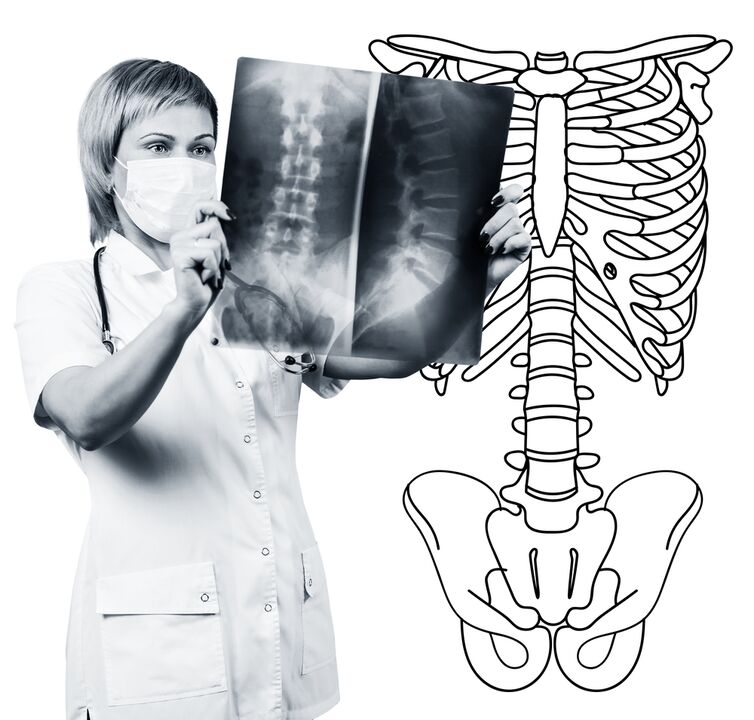
Diagnosis of osteocondrosis of the lumbar spine is based on clinical data and data on additional research methods.The most important role belongs to such methods as:
- Radiography of the lumbar spine;
- Computed tomography of the lumbar spine;
- Magnetic resonance tomography of the lumbar spine.
Radiography of the lumbar spine is necessarily performed in 2 mutually perpendicular projections-the right back and side.Such images allow you to see the shape, contours and structure of the spines, height and shape of the intervertebral disks, the abnormalities of the spine and natural bends.To show the intervertebral joints and intervertebral holes, radiograms are produced in oblique projections.To identify the pathological mobility of individual lumbar segments (which is a sign of osteocondrosis), radiography is performed under the conditions of functional experiment, that is, in flexion and expansion of the spine.Normally, you can clearly see the change at the height of the intervertebral discs in the front or rear sections in accordance with the direction of the body slope, with osteocondrosis due to the functional block of one of the segments, the height of the disk does not change when bent or extend.With pathological mobility, the displacement of the vertebrae is determined forward or backwards.The most important X -Ray signs of osteochondrosis include narrowing of the intervertebral gap, pathological mobility and displacement of vertebral bodies, deposition of salts in the disk tissue (calcification), the formation of regional growth of vertebral bodies, compaction of vertebrae at the boundary of the affected disk (subchondral sclerose).Radiography of the lumbar spine is a routine for research that gradually loses its importance based on the active implementation of new and more informative research methods (CT and MRI).Radiography over the lumbar department is used today as a screening diagnostic method.
CT of the lumbar spine is also performed using X -Ray radiation, but the radial load on the body is much smaller than with X -Ray.The study is performed on the table on a special device - a computer tomographer, it is absolutely painless.The resulting images are processed using a computer and allows you to see significantly more structures than with the spine radiography.
MRI is a method where electromagnetic radiation is used to create images.The study is also performed in the position of lying on the table, which calls into the Tomograph's Chamber.MRI is harmless and painless.
CT or MRI of the lumbar spine allows you to see all the structures of the spine, carefully examine the intervertebral disks (and the jacket and fibrous ring) and the intervertebral holes, the contents of the spinal cord duct.Even an easy projection of the intervertebral disk will not go unnoticed.These methods (especially MRI) allow you to determine the direction of disk hernia, if any, the degree of compression of nerve roots, spinal cord.Thus, these research methods are much more informative in the diagnosis of osteochondrosis of the lumbar spine than radiography.In addition, they allow you to diagnose not only osteochondrosis but also other diseases (tumors, circulation disorders in the spinal cord, abscesses, congenital defects in the spine and spinal cord), which is important during the differential diagnosis of causes of back pain.
Osteocondrosis in the lumbar spine is a disease that most often causes back pain.It is actually the destruction of the intervertebral disks.Due to osteocondrosis of the lumbar spine, a person often loses work capacity as the disease in addition to pain can lead to a violation of the spine, inability to sit, stand and walk.The symptoms of this disease are not -specific and require additional research methods to accurately confirm the diagnosis.The most informative and safe in modern methods for diagnosing osteocondrosis is MRI of the spine.